Medical Services
The Largest online database of patient reviews for doctors, facilities and online Appointment.

Pulmonary valve stenosis
(PVS) is a congenital heart defect where the pulmonary
valve leaflets thicken or fuse, narrowing the valve opening and restricting
blood flow from the right ventricle to the pulmonary arteries. This
forces the right ventricle to pump harder, potentially leading to
hypertrophy over time.
Most cases are detected in
infancy via a heart murmur during routine exams, though mild forms may remain
asymptomatic until adulthood. Symptoms in moderate-to-severe PVS include
fatigue, shortness of breath, chest pain, fainting (especially
post-exercise), cyanosis, poor weight gain in infants, and abdominal
distention. Rarely, it is associated with syndromes like Noonan or Tetralogy of
Fallot.
Diagnosis involves
echocardiography to assess valve gradient and severity, with ECG and cardiac
catheterization for confirmation. Mild cases require monitoring;
moderate-severe ones need intervention.
Primary treatment is
percutaneous balloon valvuloplasty, inflating a balloon to stretch the valve,
often curative. Severe cases may require surgical valvotomy or valve
replacement. Medications like prostaglandins, diuretics, or antiarrhythmics
support associated defects. Post-treatment, regular follow-up prevents
recurrence.
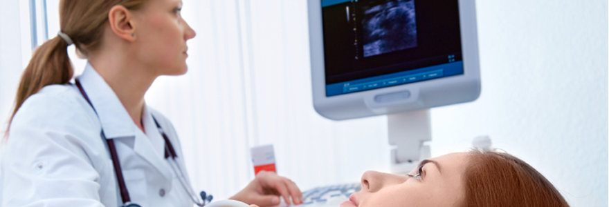
This illustration depicts normal versus stenotic pulmonary valve anatomy,
highlighting restricted outflow.